 Anatomy
The space is fringed by the membrane bone and visceral pleurae. The pleura covers the inner surface of the bodily cavity, together with the bodily cavity, diaphragm, and ribs. The pleura envelops all respiratory organ surfaces, together with the interlobar fissures. the correct and left serous membrane areas square measure separated by the bodily cavity.
The space plays a very important role in respiration by coupling the movement of the chest wall therewith of the lungs in a pair of ways in which. First, a relative vacuum within the area keeps the visceral and membrane bone pleurae in shut proximity. Second, the tiny volume of serous membrane fluid, that has been calculated at zero.13 mL/kg of weight underneath traditional circumstances, is a lubricating substance to facilitate movement of the serous membrane surfaces against one another within the course of respirations.[2] This little volume of fluid is maintained through the balance of hydraulics and oncotic pressure and humour drain, a disturbance of which can result in pathology.[3]
Etiology
The normal space contains roughly one cc of fluid, representing the balance between (1) hydraulics and oncotic forces within the visceral and membrane bone serous membrane vessels and (2) in depth humour drain. serous membrane effusions result from disruption of this balance.
Pleural effusion is associate degree indicator of associate degree underlying illness method which will be pneumonic or nonpulmonary in origin and will be acute or chronic.[4, 5] though the etiologic spectrum of serous membrane effusion is in depth, most serous membrane effusions square measure caused by symptom heart disease, pneumonia, malignancy, or embolism. the subsequent mechanisms play a task within the formation of serous membrane effusion:
- Altered porosity of the serous membrane membranes (eg, inflammation, malignancy, pneumonic embolus)
- Reduction in intravascular oncotic pressure (eg, hypoalbuminemia, cirrhosis)
- inflated capillary porosity or tube-shaped structure disruption (eg, trauma, malignancy, inflammation, infection, pneumonic pathology, drug hypersensitivity, uremia, pancreatitis)
- inflated capillary hydraulics pressure within the general and/or circulation (eg, symptom heart disease, superior venous blood vessel syndrome)
- Reduction of pressure within the space, preventing full respiratory organ enlargement (eg, in depth pathology, mesothelioma)
- minimized humour drain or complete blockage, together with lymphatic vessel obstruction or rupture (eg, malignancy, trauma)
- inflated serosa fluid, with migration across the diaphragm via the lymphatics or structural defect (eg, cirrhosis, serosa dialysis)
- Movement of fluid from pneumonic swelling across the pleura
- Persistent increase in serous membrane fluid oncotic pressure from associate degree existing serous membrane effusion, inflicting additional fluid accumulation
Pleural effusions square measure typically classified as transudates or exudates, supported the mechanism of fluid formation and serosa fluid chemistry. Transudates result from AN imbalance in oncotic and hydraulics pressures, whereas exudates square measure the results of inflammation of the serosa or reduced humour evacuation. In some cases, the serosa fluid could have a mixture of transudative and exudative characteristics.
Transudates
Transudates ar sometimes ultrafiltrates of plasma within the serous membrane because of imbalance in fluid mechanics and oncotic forces within the chest. However, they'll even be caused by the movement of fluid from serous membrane areas or by induced infusion into the space from misplaced or migrated central blood vessel catheters or gavage tubes. Transudates ar caused by atiny low, outlined cluster of etiologies, together with the following:
symptom failure
- cirrhosis of the liver (hepatic hydrothorax)
- pathology - which can flow from to malignancy or embolism
- Hypoalbuminemia
- syndrome
- serous membrane chemical analysis
- Myxedema
- Constrictive carditis
- Urinothorax - sometimes because of impeding pathology
- body fluid (CSF) leaks to the serous membrane - typically within the setting of ventriculopleural shunting or of trauma or surgery to the body part spine
- Duropleural fistula - Rare, however could also be a complication of medulla spinalis surgery
- Extravascular migration of central blood vessel catheter[7]
- Glycinothorax - A rare complication of bladder irrigation with one.5% glycine resolution following urologic surgery
Exudates
Exudates ar created by a spread of inflammatory conditions and infrequently need additional in depth analysis and treatment than transudates. Exudates arise from serosa or respiratory organ inflammation, impaired body fluid evacuation of the space, transdiaphragmatic movement of inflammatory fluid from the serous membrane area, altered permeableness of serosa membranes, and accumulated capillary wall permeableness or vascular disruption. serosa membranes ar concerned within the pathologic process of the fluid formation. permeableness of serosa capillaries to proteins is high, leading to associate degree elevated supermolecule content.
The additional common causes of exudates embody the following:
- Parapneumonic causes[8]
- Malignancy (most usually, respiratory organ or carcinoma, lymphoma, leukemia; less usually, sex gland cancer, abdomen cancer, sarcomas, melanoma)[9]
- embolism
- Collagen-vascular conditions (rheumatoid inflammatory disease, general lupus erythematosus[10] )
- infectious disease (TB)
- inflammation
- Trauma
- Postcardiac injury syndrome
- passageway perforation
- Radiation pleuritis
- pathology
- zymosis
- duct gland pseudocyst
- Intra-abdominal symptom
- Status-post arterial blood vessel bypass graft surgery
- serosa malady
- Meigs syndrome (benign girdle growth with associated pathology and serosa effusion)
- sex gland hyperstimulation syndrome
- Drug-induced serosa malady (see Pneumotox On Line for an intensive list of medication that may cause serosa effusion)
- Asbestos-related serosa malady
- Yellow nail syndrome (yellow nails, lymphedema, serosa effusions)
- Uremia
- cornered respiratory organ (localized serosa scarring with the formation of a protein peel prevents incomplete respiratory organ enlargement, from time to time resulting in serosa effusion)
- Chylothorax (acute health problem with elevated triglycerides in serosa fluid)
- Pseudochylothorax (chronic condition with elevated steroid alcohol in serosa fluid)
- Fistula (ventriculopleural, biliopleural, gastropleural)
A detailed medical record ought to be obtained from all patients presenting with a serosa effusion, as this might facilitate to determine the etiology. for instance, a history of chronic liver disease or alcoholism with cirrhosis of the liver suggests internal organ congestion or alcohol-induced redness with effusion. Recent trauma or surgery to the body part spine raises the likelihood of a CSF leak. The patient ought to be asked a couple of history of cancer, even remote, as malignant serosa effusions will develop a few years once initial identification.
An activity history ought to even be obtained, as well as potential amphibole exposure, that might dispose the patient to carcinoma or amphibole serosa effusion. The patient ought to even be asked regarding medications they're taking.[4]
The clinical manifestations of serosa effusion square measure variable and sometimes square measure associated with the underlying illness method. the foremost usually associated symptoms square measure progressive symptom, cough, and pleuritic hurting.
Dyspnea
Dyspnea is that the most typical symptom related to serosa effusion and is expounded a lot of to distortion of the diaphragm and chest wall throughout respiration than to hypoxemia. In several patients, drain of serosa fluid alleviates symptoms despite restricted improvement in gas exchange. drain of serosa fluid may additionally permit the underlying illness to be recognized on repeat chest radiographs. Note that symptom is also caused by the condition manufacturing the serosa effusion, like underlying intrinsic respiratory organ or heart condition, obstructing endobronchial lesions, or diaphragmatic dysfunction, instead of by the effusion itself.
Cough
Cough in patients with serosa effusion is commonly gentle and unproductive. a lot of severe cough or the assembly of infected or bloody humour suggests AN underlying respiratory disease or endobronchial lesion.
Chest pain
The presence of hurting, which ends from serosa irritation, raises the chance of AN exudative etiology, like serosa infection, carcinoma, or pneumonic pathology.[18]
Pain is also gentle or severe. it's generally delineate as sharp or stabbing and is exacerbated with deep inspiration. Pain is also localized to the chest wall or cited the ipsilateral shoulder or higher abdomen, actually because of diaphragmatic involvement. Pain usually diminishes in intensity because the serosa effusion will increase in size.
Additional symptoms
Other symptoms in association with serous membrane effusions might recommend the underlying unwellness method. Increasing lower extremity swelling, orthopnea, and attack nocturnal dyspnoea might all occur with symptom heart condition.
Night sweats, fever, hemoptysis, and weight loss ought to recommend TB. haemoptysis additionally raises the likelihood of malignancy, different endotracheal or endobronchial pathology, or pulmonic pathology. associate degree acute feverish episode, infected bodily fluid production, associate degreed pleuritic hurting might occur in patients with an effusion related to respiratory disorder.
Physical Examination
Physical findings in serosa effusion square measure variable and rely on the amount of the effusion. Generally, there are not any physical findings for effusions smaller than three hundred mil. With effusions larger than three hundred mil, findings could embody the following:
- Dullness to percussion, remittent tactile fremitus, and asymmetrical chest growth, with diminished or delayed growth on the aspect of the effusion, square measure the foremost reliable physical findings of serosa effusion.[19, 20]
- Mediastinal shift off from the effusion - this can be discovered with effusions of larger than a thousand mL; displacement of the trachea Associate in Nursingd cavum toward the aspect of the effusion is a crucial clue to obstruction of a body part cartilaginous tube by an endobronchial lesion, which might flow from to malignancy or, less normally, to a benign cause, like a far off body.
- Diminished or quiet breath sounds
- Egophony ("e" to "a" changes) at the foremost superior side of the serosa effusion
- serosa friction rub
Peripheral puffiness, distended neck veins, and S3 gallop counsel symptom coronary failure. puffiness can also be a manifestation of nephrotic syndrome; serosa disease; or, combined with yellow nails, the yellow nail syndrome.
connective tissue changes with pathology counsel disease
pathology or a palpable mass suggests malignancy.[4]
Differential Diagnoses
- CBRNE - Q Fever
- Congestive Heart Failure and Pulmonary Edema
- Diaphragmatic Injuries in Emergency Medicine
- Esophageal Rupture and Tears in Emergency Medicine
- Hypothyroidism and Myxedema Coma in Emergency Medicine
- Neoplasms, Lung
- Pancreatitis
- Rheumatoid Arthritis
Thoracentesis ought to be performed for brand spanking new and unexplained serous membrane effusions once adequate fluid is gift to permit a secure procedure. Observation of serous membrane effusion is affordable once benign etiologies area unit probably, as within the setting of open symptom heart disease, infectious agent inflammatory disease, or recent body part or abdominal surgery.
Laboratory testing helps to tell apart serous membrane fluid transudates from exudates; but, bound kinds of exudative serous membrane effusions may be suspected just by observant the gross characteristics of the fluid obtained throughout thoracentesis. Note the following:
- candidly infected fluid indicates Associate in Nursing inflammatory disease
- A putrid odor suggests Associate in Nursing anaerobic inflammatory disease
- A milky, iridescent fluid suggests a chylothorax, ensuing most frequently from bodily fluid obstruction by malignancy or lymphatic vessel injury by trauma or operation
- Grossly bloody fluid could result from trauma, malignancy, postpericardiotomy syndrome, or asbestos-related effusion and indicates the requirement for a spun hematocrit check of the sample; a serous membrane fluid hematocrit level of over five hundredth of the peripheral hematocrit level defines a congestion, which frequently needs tube thoracostomy
Normal serous membrane fluid has the subsequent characteristics:
- Clear ultrafiltrate of plasma that originates from the pleura
- A pH of 7.60-7.64
- macromolecule content of but two (1-2 g/dL)
- Fewer than a thousand white blood cells (WBCs) per metric capacity unit
- aldohexose content like that of plasma
- nurse dehydrogenase (LDH) but five hundredth of plasma
Chest Radiography
Effusions of over a hundred seventy five metric capacity unit square measure typically apparent as blunting of the costophrenic angle on upright posteroanterior chest radiographs. On supine chest radiographs, that square measure ordinarily employed in the medical care setting, moderate to massive serous membrane effusions might seem as the same increase in density contact the lower respiratory organ fields. Apparent elevation of the hemidiaphragm, lateral displacement of the dome of the diaphragm, or inflated distance between the apparent left hemidiaphragm and also the viscus bubble suggests subpulmonic effusions. (See the photographs below.)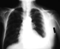
Diagnostic Thoracentesis
Perform diagnostic thoracentesis if the etiology of the effusion is unclear or if the plausible reason behind the effusion doesn't reply to medical aid obviously. serous membrane effusions don't need thoracentesis if they're too tiny to securely aspirate or, in clinically stable patients, if their presence will be explained by underlying symptom heart condition (especially bilateral effusions) or by recent body part or abdominal surgery.
Depending on the clinician’s expertise, a pulmonologist will be consulted for help with unsound diagnostic thoracentesis.
Contraindications
Relative contraindications to diagnostic thoracentesis embrace atiny low volume of fluid (< one cm thickness on a lateral posture film), trauma sensitivity or general medical aid, mechanical ventilation, and body covering unwellness over the planned puncture web site. Mechanical ventilation with positive end-expiratory pressure doesn't increase the danger of abnormality when thoracentesis, however it will increase the chance of severe complications (tension abnormality or persistent bronchopleural fistula) if the respiratory organ is pierced.
Complications of diagnostic thoracentesis embrace pain at the puncture web site, body covering or internal trauma, abnormality, empyema, and spleen/liver puncture. abnormality complicates roughly 12-30% of thoracenteses however needs treatment with a chest tube in but five-hitter of cases. Use of needles larger than twenty gauge will increase the danger of a abnormality complicating the thoracentesis. additionally, important chronic hindering or fibrotic respiratory organ unwellness will increase the danger of a symptomatic abnormality complicating the thoracentesis.
Procedure
In patients with giant, freely flowing effusions and no relative contraindications to thoracentesis, diagnostic thoracentesis will typically be performed safely, with the puncture web site ab initio chosen supported the chest exposure and set 1-2 rib interspaces below the amount of dullness to percussion on physical examination. In different things, imaging or chest CT scanning ought to be accustomed guide thoracentesis.
After the positioning is disinfected with antiseptic (preferred) or povidone/iodine (no longer recommended) resolution and sterile drapes ar placed, drug the skin, periosteum, and pleura with a hundred and twenty fifth Xylocaine through a 25-gauge needle. If serosa fluid isn't obtained with the shorter 25-gauge needle, continue anesthetizing with a one.5-inch, 22-gauge needle. For patients with larger amounts of hypodermic tissue, a 3.5-inch, 22-gauge spinal needle with inner style removed may be accustomed drug the deeper tissues and notice the effusion.
Confirm the proper location for thoracentesis by aspirating serosa fluid through the 25- or 22-gauge needle before introducing larger-bore thoracentesis needles or catheters. If serosa fluid isn't simply aspirated, stop the procedure and use imaging or chest CT scanning to guide thoracentesis.
When attainable, patients ought to sit upright for thoracentesis. Patients shouldn't lean forward, as a result of this causes serosa fluid to maneuver to the anterior costophrenic area and will increase the chance of puncture of the liver or spleen. For debilitated and oxygenated patients UN agency cannot sit upright, acquire serosa fluid by puncturing over the eighth rib at the midaxillar to posterior axillary line. In such patients, imaging is also needed to guide thoracentesis.
Supplemental gas is usually administered throughout thoracentesis to offset hypoxemia created by changes in ventilation-perfusion relationships as fluid is removed and to facilitate biological process of serosa air if abnormal condition complicates the procedure.
The frequency of complications from thoracentesis is lower once a experienced practician performs the procedure and once ultrasonographic steerage is employed.[33] Consequently, a talented and intimate practician ought to perform thoracentesis in patients UN agency have the next risk of complications or relative contraindications for thoracentesis and in patients UN agency cannot sit upright.
Postprocedure breath chest radiographs to exclude abnormal condition aren't required in symptomless patients once uncomplicated procedures (single needle pass while not aspiration of air). However, postprocedure breath chest radiographs ar counseled to determine a replacement baseline for patients possible to own perennial symptomatic effusions.
Medication outline
Pharmacologic management of serous membrane effusion depends on the condition’s etiology. for instance, medical management includes nitrates and diuretics for symptom coronary failure and pneumonic puffiness, antibiotics for parapneumonic effusion and inflammatory disease, and medical aid for embolism.
In patients with parapneumonic effusions, empyemas, and effusions related to muscle system perforation and intra-abdominal abscesses, antibiotics ought to be administered early once these conditions square measure suspected.
Antibiotic choice ought to be supported the suspected motive microorganisms and therefore the overall clinical image. issues embrace the patient's age, comorbidities, length of the health problem, setting (community vs nursing home), and native organism sensitivities. varied effective single agents and combination antimicrobial therapies exist. Coverage ought to typically embrace anaerobic organisms. choices could embrace clindamycin, extended-spectrum penicillins, and imipenem. looking on the patient's clinical condition, communicable disease consultation could also be acceptable.
Particular attention should run to potential drug interactions, adverse effects, and preexistent conditions.


No comments:
Post a Comment